Rigid Ureteroscope: Uses, Benefits, and Medical Importance
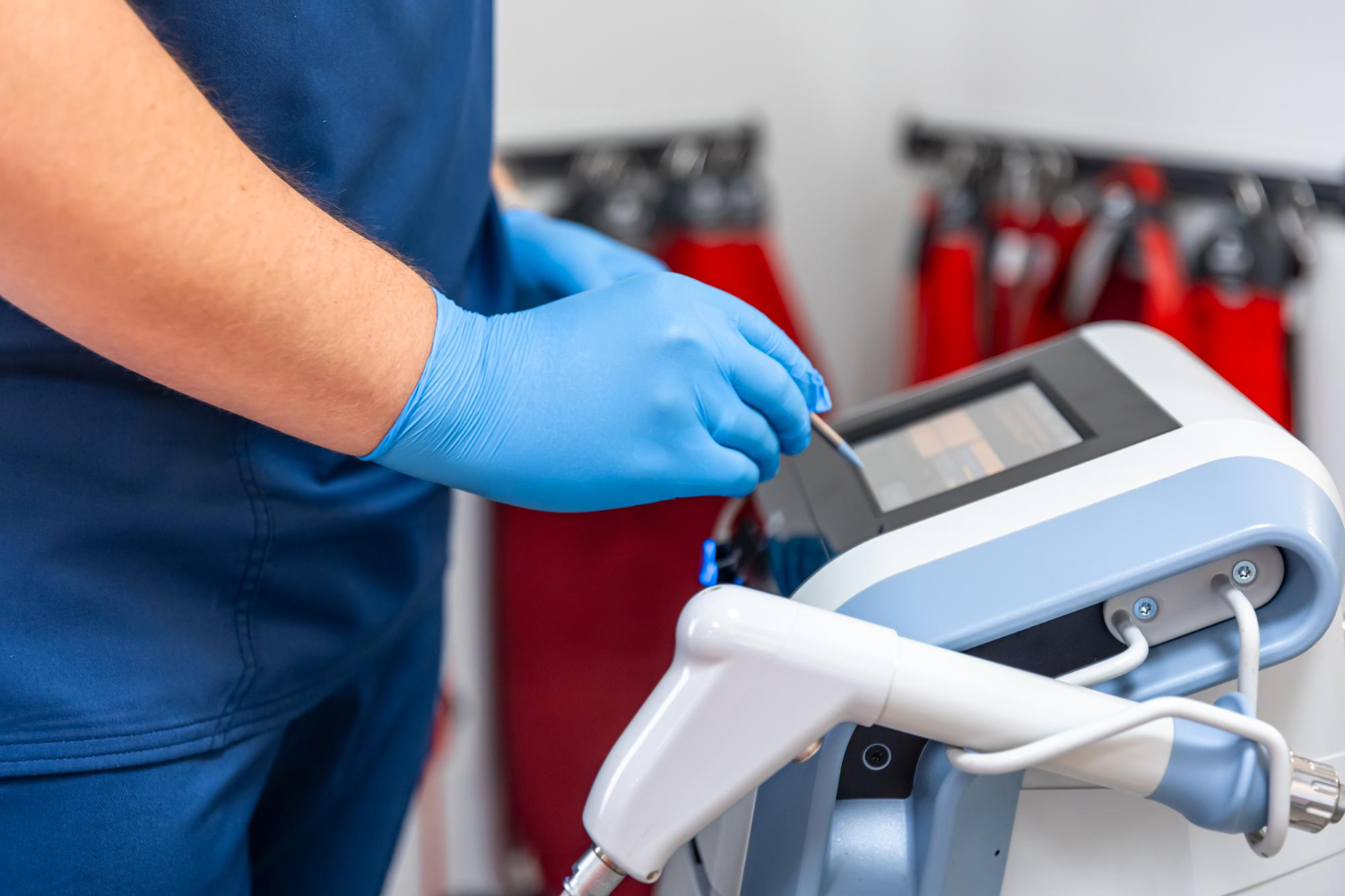
A rigid ureteroscope is a medical instrument used by urologists to examine and treat problems inside the urinary tract. It is a thin, straight tube with a light and camera attached at the end. Doctors insert it through the urethra and bladder into the ureter to see stones, tumors, or blockages. This device plays an important role in modern urology because it allows treatment without large cuts or open surgery.
What Is a Rigid Ureteroscope?
A rigid ureteroscope is a straight, non-flexible scope designed to access the lower and middle part of the ureter. The ureter is the tube that carries urine from the kidney to the bladder. This instrument helps doctors see inside the urinary system clearly. It is commonly made from stainless steel and contains fiber-optic lighting for better visibility.
Unlike flexible scopes, this device does not bend. Because of its straight design, it provides clear imaging and better control during certain procedures. It is mostly used when the stone or problem is located in the lower ureter.
Why Doctors Use It
Urologists use a rigid ureteroscope mainly to diagnose and treat ureteral stones. Kidney stones can move into the ureter and cause severe pain, infection, or blockage. When stones are too large to pass naturally, doctors remove or break them using this instrument.
It is also used to:
-
Remove small tumors
-
Treat ureteral strictures (narrowing)
-
Take biopsy samples
-
Place ureteral stents
-
Examine bleeding causes
This tool helps avoid open surgery in many cases, which means faster recovery and less pain for patients.
How the Procedure Is Done
The procedure using a rigid ureteroscope is called ureteroscopy. It is usually done under general or spinal anesthesia. The doctor gently inserts the scope through the urethra into the bladder and then into the ureter.
If a stone is found, it can be removed using small forceps or broken into pieces with a laser. After treatment, a temporary stent may be placed to help urine flow properly.
The entire procedure usually takes 30 to 90 minutes depending on the case. Most patients can go home the same day.
Main Advantages
There are many benefits of using a rigid ureteroscope:
1. Minimally Invasive
There are no large cuts on the body. The scope enters through natural urinary passages.
2. High Success Rate
It is very effective for lower ureter stones.
3. Short Recovery Time
Most patients recover quickly and return to daily activities within a few days.
4. Clear Visibility
The straight design provides stable imaging.
5. Cost-Effective
Compared to some flexible systems, it is more affordable.
Because of these advantages, it is widely used in hospitals and urology centers.
Rigid vs Flexible Ureteroscope
Both rigid and flexible scopes are used in urology, but they serve different purposes.
-
Rigid scopes are best for lower ureter stones.
-
Flexible scopes are better for stones located inside the kidney.
Rigid instruments are usually stronger and allow better control during stone removal. Flexible ones can bend and reach upper areas of the kidney.
Doctors choose the type depending on stone location, size, and patient condition.
Risks and Safety
Like any medical procedure, ureteroscopy has some risks. However, serious problems are rare when performed by experienced doctors.
Possible risks include:
-
Mild bleeding
-
Urinary infection
-
Temporary discomfort while urinating
-
Ureter injury (rare)
Most side effects are mild and improve within a few days. Drinking plenty of water after the procedure helps recovery.
Technological Improvements
Modern rigid ureteroscope models have improved over time. New designs include better lighting systems and smaller diameters. Smaller scopes reduce discomfort and lower the risk of injury.
High-definition cameras now provide clear images, allowing doctors to see even small stones or lesions. Some devices also include irrigation channels to maintain a clear view during procedures.
These improvements have made ureteroscopy safer and more effective.
Who Needs This Procedure?
Patients who may need treatment using a rigid ureteroscope include those with:
-
Persistent ureter stones
-
Severe flank pain
-
Blood in urine
-
Urinary blockage
-
Repeated urinary infections
Doctors usually confirm the need through ultrasound, CT scan, or X-ray imaging.
If stones are too big to pass naturally or cause complications, ureteroscopy is recommended.
Preparation Before Procedure
Before undergoing ureteroscopy, patients may need:
-
Blood tests
-
Urine tests
-
Imaging scans
-
Fasting for several hours
Doctors may prescribe antibiotics to prevent infection. It is important to inform the doctor about any medications being taken.
Recovery After Procedure
Recovery is usually quick. Patients may feel mild burning during urination for one or two days. Some may notice small blood traces in urine, which is normal.
If a stent is placed, mild discomfort may continue until it is removed. Doctors usually remove the stent after one to two weeks.
Patients are advised to:
-
Drink plenty of water
-
Avoid heavy lifting
-
Take prescribed medicines
-
Report fever or severe pain immediately
Following medical advice ensures smooth healing.
Importance in Modern Urology
The rigid ureteroscope has changed how doctors treat urinary stones. In the past, open surgery was common for stone removal. Today, minimally invasive methods are preferred.
This instrument helps reduce hospital stay and medical costs. It also lowers the risk of complications compared to traditional surgery.
Because kidney and ureter stones are becoming more common worldwide due to lifestyle and dietary habits, ureteroscopy remains an essential treatment option.
Conclusion
A rigid ureteroscope is a vital tool in urology used to diagnose and treat ureter problems, especially stones. It provides a safe, effective, and minimally invasive solution for patients suffering from urinary blockages. With modern technology and improved design, it continues to offer high success rates and fast recovery.
This device plays an important role in reducing pain, avoiding open surgery, and improving patient outcomes. As medical technology advances, its performance and safety will continue to improve, making it an essential instrument in hospitals around the world.